


The Biochemistry and Bioelectricity of Injured Tissue
The Biochemistry of Injury
Following injury, damaged cells in injured tissue immediately “amp up” as they kick into action, initiating multiple self-repair mechanisms.
They begin discharging certain bio-chemical substances, (such as arachidonic acid, a component of the phospholipid structure of the cell membrane itself) from wounds into their immediate surroundings. From this, prostaglandins are synthesized, triggering a cascade of reactions resulting in the release of histamines and bradykinins (amino acids) – which can stimulate pain receptors as well as partake in the inflammatory response.
Blood flows into the area. Fluids accumulate; swelling occurs. Heat rises. Redness appears. In time, if the cells are unable to complete their repair job, these substances remain in the tissue and are responsible for infections, plus lingering (chronic) inflammation, producing a persistent painful sensation and associated pathological conditions.
The Bioelectricity of Injury
When acute cellular repair work is underway, while chemicals, such as histamine, etc., are building up in the tissue surrounding an injury, there is an initial spike in electrical activity in all involved cells; and increased conductivity of currents, because fluids are being produced and carried to the site and fluids are highly conductive (like electricity through water).
After a time, cellular energy production slows down, eventually becoming depleted, especially if the system is weak and tired in general. As circulation decreases, remaining substances clog the tissue, producing a measurable decrease in electrical conductivity (an increase in tissue resistance).
These differences in the bio-electricity are measurable using sensitive monitoring devices.
Here are familiar ways we measure bio-electricity:
- brainwaves move across the brain (EEG)
- skin resistance (GSR)
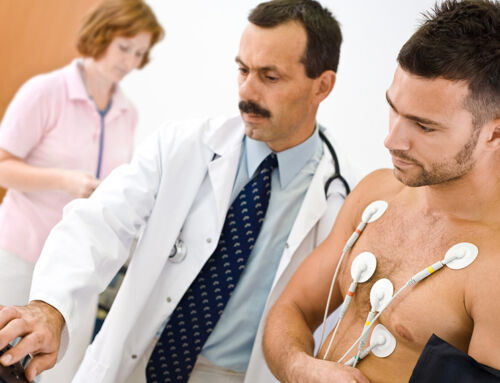
- heart activity (EKG)
- muscle tissue conductance (EMG)
It is evident that every cell in the body generates tiny electrical currents. These well-known biofeedback devices can tell us about the bio-electricity of our heart, brain, skin, and muscles. When electrical currents are blocked in the brain or heart, fatal results can follow.
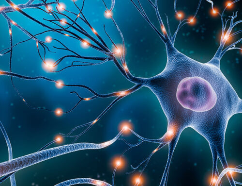
Nerve conduction biofeedback is not as well-known. Nerve Conduction Studies (NCS) and Nerve Conduction Velocity (NCV) are measurements of the electrical activity of nerve cells, or neurons, which are highly excitable. When stimulated, they produce tiny traveling waves of electricity – nerve signals, or impulses. These pass along to other neurons, eliciting similar responses from them. Thus, miniscule waves of bio-electricity are propelled along linear nerve pathways (relaying their “spark” from neuron to neuron), throughout the Nervous System.
Advanced Biotechnology
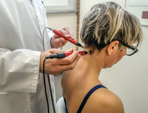
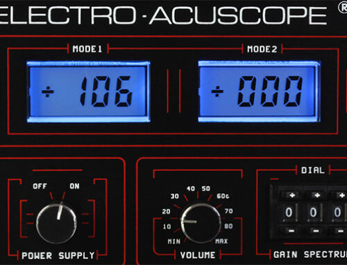
The Acuscope has built-in nerve monitoring capability. Its biofeedback component “picks up” the micro-electrical signals being generated by nerve signal conduction, miniscule currents moving through tissue.
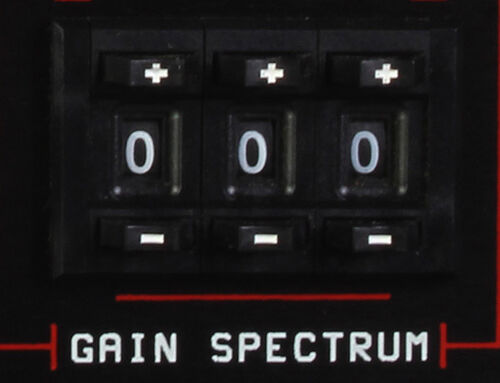
A number display reveals to the Operator exactly where tissue inflammation is located (the nerve cells are hyper-excited and nerve endings highly activated); and further shows where surrounding tissue has become depleted and discharged (sluggish, drained of “energy”/exhausted).
The Acuscope can then send in a signal to boost where a re-charge is required and/or to disperse the congestion where there is (or has been) inflammation. When the correct Frequencies of biologically-compatible microcurrents are applied to tissue in abnormal states of conductivity, homeostasis (balance) returns; the cells can finally finish their incomplete self-repair jobs, pain subsides, and old, lingering issues become resolved and disappear.
This end-result may take time, a series of treatments over days, weeks, or months (in severe conditions); however, it is a natural, non-invasive solution, frequently producing accelerated tissue repair, and is frequently responsible for resolution which may never have otherwise occurred.
The Myopulse is also monitoring biofeedback: the ability of contractile cells to do their work is measurable in terms of cellular electrical potentials (the amount of electrical charge required for a cell to produce an action e.g., its potential to fire). Over-firing muscle cells will eventually lead to tightness, knots, spasms, and can also result from pulls, sprains, strains and/or painful repetitive use syndromes. Under-firing muscle cells are evident in aging, atrophied, and weak connective tissue as well as in old injury comprised of cells which have never completely finished repairing themselves.